How a Vascular Ultrasound Can Help Diagnose Circulatory Disorders
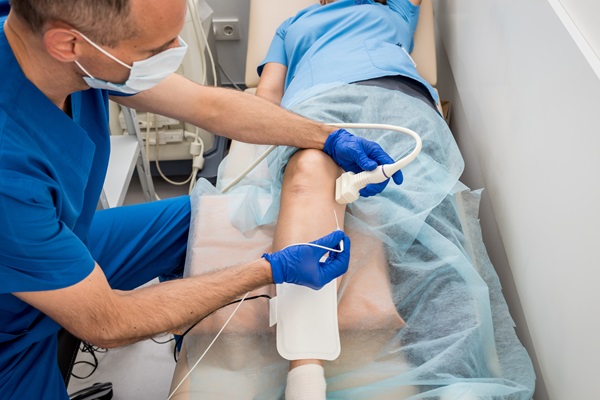
Also called a duplex study, a vascular ultrasound is a noninvasive evaluation of how much blood flows through your neck, legs, and arms. The process uses high-frequency sound waves to create pictures of soft tissue and blood vessels. The ultrasound allows the physician to figure out the movement and speed of blood cells, as well as determine the blood vessels’ condition. The exam can locate blockages or issues within vessels.
Why get a vascular ultrasound?
The arteries transport oxygen-rich blood from the heart to the rest of the entire body, while oxygen-depleted blood returns via the veins to the heart for reoxygenation. This system constitutes the majority of the circulatory system or the vascular system. A medical professional might suggest a vascular ultrasound exam for the following reasons:
- Monitor blood flow to certain organs and tissues
- Identify changes in organ or tissue appearance
- Find abnormal masses like tumors
- Find any obstructions, anomalies, and even narrowing of blood vessels (vascular stenosis)
- Measure the rate of blood flow
- Detect blood clots
- Find an enlarged blood vessel
- Evaluate varicose veins
- Determine whether an angioplasty is required
- Determine whether a surgery is successful
The vascular ultrasound process
Ultrasound imaging utilizes sonar principles. If sound waves hit an object, it bounces back or even echoes. Measuring these echo waves can help estimate the distance of an object, its consistency, shape, and size. It can also show if the object is solid or fluid-filled.
A transducer transmits the audio waves and records the echoed waves in an ultrasound exam. The technologist will press the transducer over the skin, sending tiny pulses of unintelligible, high-frequency sound into the body. When the sound waves reflect off internal organs, fluids, and tissues, the sensitive receiver of the transducer records subtle changes in pitch and direction.
These signature waves show on a monitor in real time and are evaluated instantly by a computer. The technologist will take one or more frames of the moving photographs as still images or short video loops.
Doppler ultrasound can determine how blood cells move and their speeds. The motion of blood cells triggers an alteration of the pitch of reflected sound waves, called the Doppler effect. The computer records the sounds and also produces charts or color photographs that show how the blood flows through the vessels.
What to expect
During the procedure, patients will lie face-up on an adjustable examination table. The professional might adjust the patient’s position to get a better image. They will then apply a clear water-based gel to the area being checked. This helps the transducer stick with the human body. Additionally, it will also prevent air pockets from forming between the transducer and the skin, which can prevent sound waves from entering the body. The radiologist will sweep over the area at different points. They can also aim the sound beam from another location for a different perspective.
After the examination, the technologist might ask the patient to wait while they review the ultrasound pictures. An ultrasound examination will typically take around 30 to 45 minutes to complete. Sometimes, the exam might take longer, depending on the complexity of the situation.
In conclusion
If you think you might have a circulation issue, you should go for an examination with a vascular doctor as soon as possible. Circulation issues and blockages can result in serious problems including a heart attack, pulmonary embolism, or stroke if not detected early enough. The doctor will perform a vascular ultrasound to assess the situation and recommend the course of action.
Get more information here: https://visoc.org or call Vascular & Interventional Specialists of Orange County at (714) 598-1194.
Check out what others are saying about our services on Yelp: Vascular Ultrasound in Orange, CA.
Related Posts
Leg swelling is a common symptom that can range from mild to severe and has many causes. Sometimes, the cause is temporary and of mild to moderate concern, but leg swelling may point to a more serious health condition in other cases. Learning more about leg swelling helps people know when to seek help from…
Hemorrhoid treatment options vary depending on severity and symptoms. There are many effective ways to manage hemorrhoids, such as through lifestyle changes, over-the-counter remedies, or more advanced medical procedures. Finding the right treatment can make a significant difference in achieving lasting relief.Hemorrhoids are swollen veins located in or around the anus and rectum, with internal…
Most uterine fibroids are noncancerous, and many patients do not realize they have them. This often leaves patients confused about what could have caused their fibroids, while they also wonder what exactly fibroids are. An OB/GYN can provide clarification on a patient’s unique condition. However, in the meantime, an overview may help.Fibroids are muscular tumors…
Curious about what varicose vein treatment from a cardiologist entails? Read on to learn more. Varicose vein treatment can significantly improve your appearance as well as your life. Varicose veins are enlarged, ropey veins that typically appear on the legs and feet. These oversized veins can often cause swelling, fatigue, and pain. They can also…